Breast MRI Features May Predict Second Cancer Risk in Breast Cancer Survivors
Women with a personal history of breast cancer face an elevated risk of developing a second breast cancer, particularly if they have dense breast tissue. A recent study published in Radiology, the journal of the Radiological Society of North America (RSNA), sheds light on how certain features visible on breast MRI may help predict this risk.
Breast Cancer and Second Cancer Risk
Breast cancer remains the most frequently diagnosed cancer and a leading cause of cancer-related death among women worldwide. Advances in early detection and treatment have significantly improved survival rates, but survivors are at higher risk for developing second breast cancers.
This risk is even greater for women with dense breasts, which have more fibroglandular tissue compared to fatty tissue. Dense breast tissue is an independent risk factor for breast cancer and can make detecting cancerous lesions on mammography more challenging.
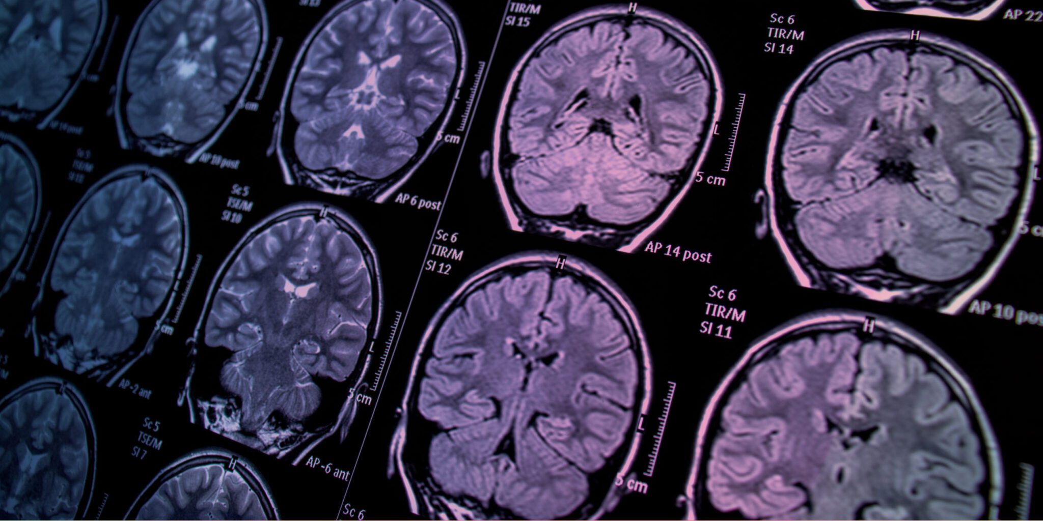
Breast MRI, a highly sensitive imaging tool, has become the preferred screening method for women with a personal history of breast cancer. Studies show that breast MRI outperforms mammography in detecting breast cancer, particularly in women with dense breasts.
What the Study Found
Led by Dr. Su Hyun Lee of Seoul National University Hospital, the study focused on a breast MRI feature known as background parenchymal enhancement (BPE). BPE refers to the brightening of breast tissue on MRI after a contrast agent is administered. This enhancement varies between individuals and reflects changes in blood supply and hormone levels in breast tissue.
The study included 2,668 women who underwent postoperative surveillance with breast MRI. Over a median follow-up of 5.8 years, 109 women developed a second breast cancer. The researchers found that women with mild, moderate, or marked BPE at their MRI scans were at a higher risk of developing a second breast cancer compared to those with minimal BPE.
Dr. Lee noted that these findings suggest BPE could reflect how breast tissue responds to cancer treatment and may serve as a predictor of modified risk after treatment.
Implications for Personalized Screening
The study highlights the potential of BPE as a tool for refining breast cancer surveillance strategies. Women with minimal BPE may no longer require annual contrast-enhanced breast MRI if no other risk factors are present, such as younger age at initial diagnosis, genetic mutations, or hormone receptor-positive tumors.
“These results could help us stratify the risk of second breast cancer and tailor surveillance strategies,” Dr. Lee said. “For instance, we might adjust the frequency and type of imaging based on BPE levels and other individual risk factors.”
Future Directions
Dr. Lee and her team plan to explore the relationship between changes in BPE—visible on preoperative and postoperative MRI—and the likelihood of second breast cancer. They anticipate that combining data from mammography, ultrasound, and MRI will lead to more personalized surveillance strategies in the future.
By integrating these findings, medical professionals may better identify women at high risk for second cancers and reduce the imaging burden for those with a lower risk profile.
This study underscores the importance of advanced imaging techniques and individualized care in managing the ongoing health of breast cancer survivors. As researchers continue to refine risk models, the goal remains clear: improving outcomes and quality of life for breast cancer patients worldwide.
Reference : Breast MRI reduces risk of second breast cancer


