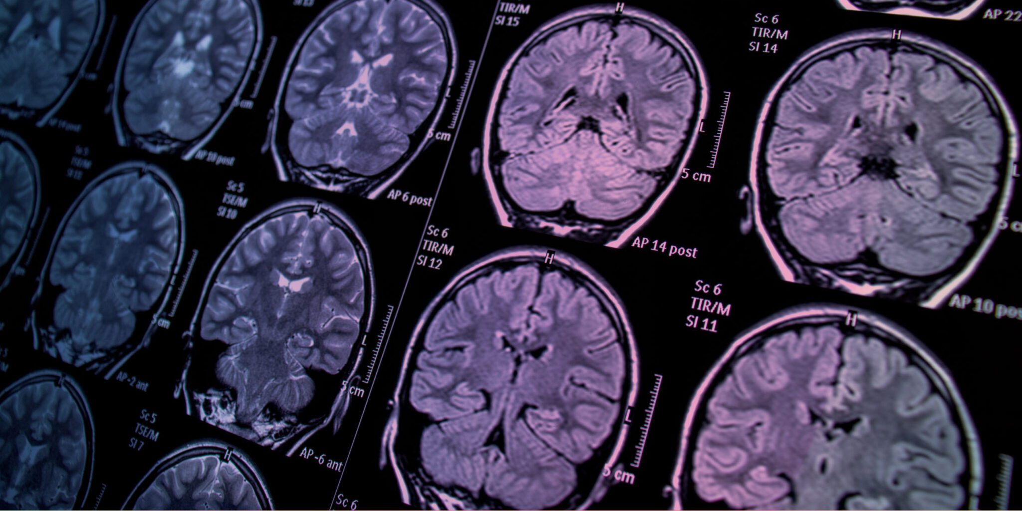

Revolutionary Medical Imaging Technology Pioneered at Rutgers University Aims to Transform Early Cancer Detection
In a groundbreaking advancement, researchers at Rutgers University are developing a state-of-the-art medical imaging technique that holds the potential to detect cancer and other diseases much earlier than existing methods. This revolutionary approach could not only expedite treatment but also significantly reduce the reliance on invasive and time-consuming biopsies.
The innovative method employs nanotechnology to illuminate small cancerous tumors and cardiovascular lesions located deep within the body. Rutgers researchers from the schools of engineering and pharmacy have reported promising results in early trials. Their pioneering work, published in the July edition of Nature Communications, recently secured a $2.2 million grant from the National Institute of Biomedical Imaging and Bioengineering, a division of the National Institutes of Health, to further advance the research.
“Our new fluorescent imaging approach not only aims to detect diseases at an earlier stage but also enables us to gather valuable insights about the diseases before performing surgeries,” explains Prabhas Moghe, the project’s lead researcher and distinguished professor of biomedical engineering and chemical and biochemical engineering at Rutgers. “I think of it as an optical biopsy.”
Addressing Key Surgical Challenges
One of the major applications of this technology is in accurately assessing whether a newly detected cancer has spread to nearby lymph nodes. This capability could allow surgeons to fully address the extent of the disease during a single operation.
“Currently, surgeons often rely on lymph node biopsies and may need to wait for results before planning a second surgery, which can lead to additional trauma, risks, and costs,” says Shridar Ganesan, associate director for Translational Science at Rutgers Cancer Institute of New Jersey and clinical advisor for the project. “This technique has the potential to streamline that process and significantly enhance patient outcomes.”
Harnessing the Power of Shortwave Infrared Light
The Rutgers imaging technology, co-developed by Richard Riman, distinguished professor of Materials Science and Engineering, uses shortwave infrared light, an alternative to the infrared light traditionally used in medical imaging. This type of light penetrates deeper into skin and tissues compared to visible or near-infrared light, providing superior imaging capabilities.
Key to this advancement are fluorescent dyes made from nanocrystals of rare-earth elements—a group of 17 metals known for their unique optical properties. Rare-earth elements, while not scarce, are challenging to mine and are increasingly in demand for applications in electronics, including smartphones, video screens, and electric vehicle components.
Earlier attempts to use shortwave infrared light for medical imaging were hindered by the toxicity of available fluorescent dyes or their inability to produce sharp images. The Rutgers team has overcome these limitations by encapsulating rare-earth nanocrystals within a shell of human serum albumin. This innovative design makes the dyes biocompatible, allowing them to distribute quickly throughout the body and concentrate at disease sites.
A Breakthrough in Precision Medicine
The researchers are further enhancing the technology by utilizing different types of rare-earth elements. Each element fluoresces in slightly distinct colors under shortwave infrared light, enabling the creation of a family of highly specific probes. These probes can identify a wide range of cancers and provide detailed information on the makeup and progression of the disease.
“This capability allows us to build a precise picture of the disease’s stage and composition,” Moghe adds.
Early Success and Future Prospects
Initial laboratory tests in mice have demonstrated that the technology can detect cancer spread on a microscopic scale earlier than traditional imaging techniques such as magnetic resonance imaging (MRI) or near-infrared imaging. By identifying diseases at an earlier stage, this method has the potential to open up new pathways for timely intervention and treatment.
The promising results of this research mark a significant step forward in the field of medical imaging. With continued advancements and clinical testing, this groundbreaking technique could soon become a vital tool in the fight against cancer and other life-threatening diseases.
Reference : Detecting cancer earlier new medical imaging technology